


Instant
Digital Pathology
Ex vivo imaging. Rapid on-site evaluation workflow.
In just 5 minutes.
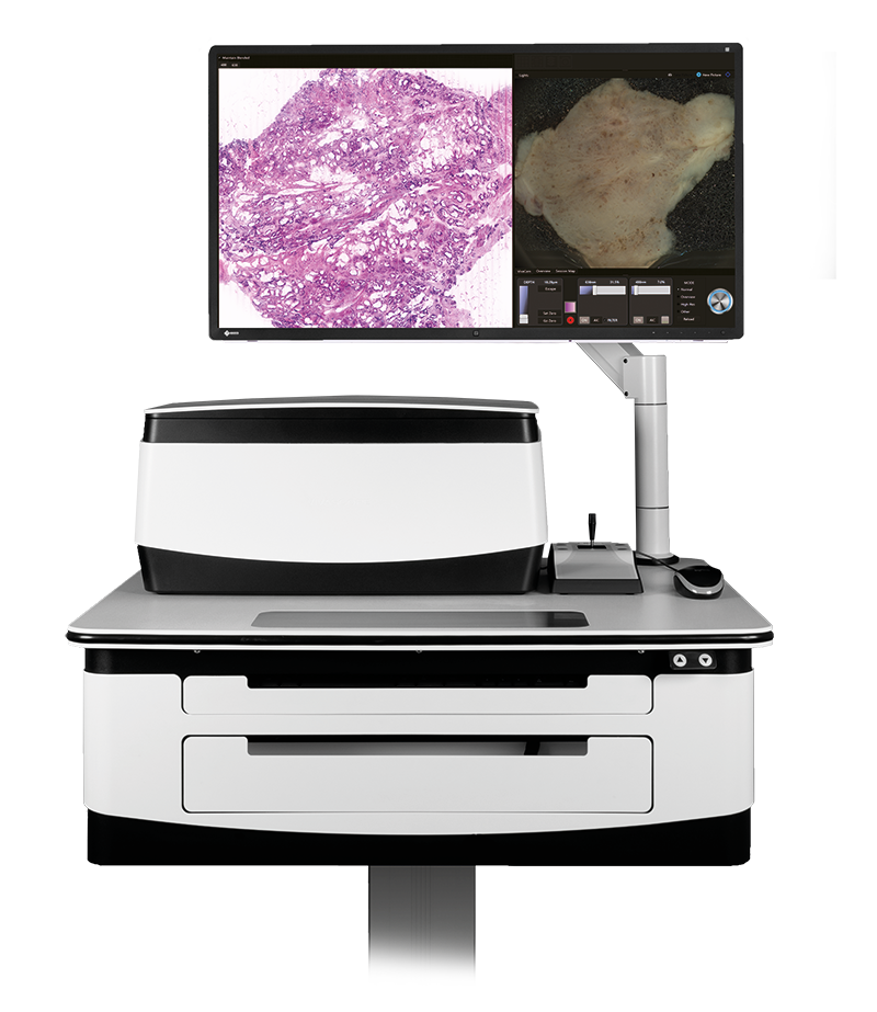
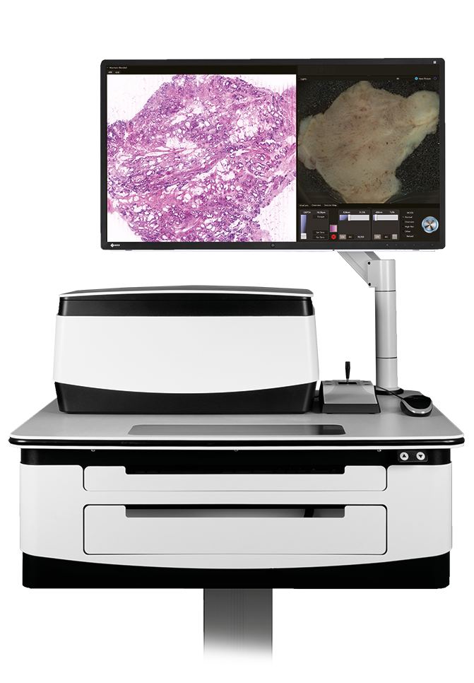
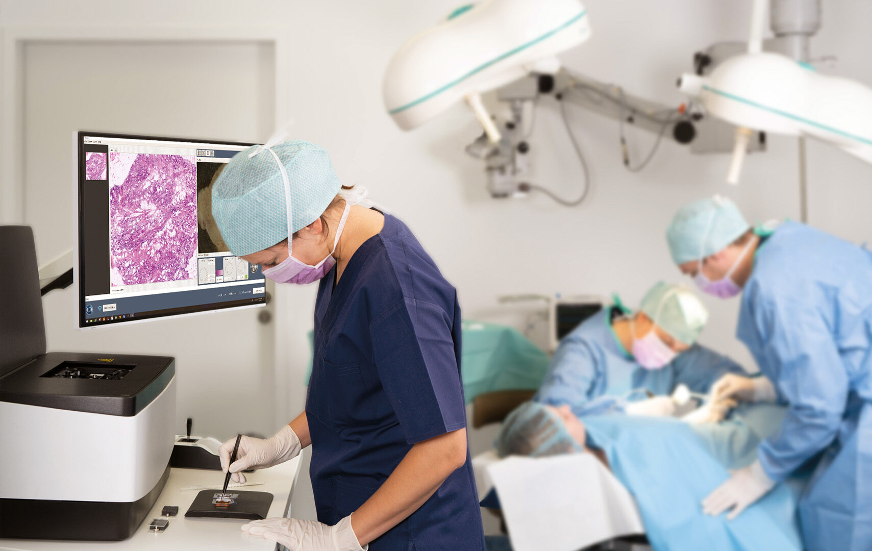
H&E-like digital images. Intraoperatively. At the beside. In just 5 minutes.
VivaScope technology is based on confocal microscopy (CLSM) and acquires images of superb optical resolution and contrast. Two lasers of different wavelengths create two distinct images, a fluorescence image and a reflectance image. Both signals are scanned simultaneously and are used to create pseudo-colored images. The device’s software uses an algorithm to translate the acquired image information into colors that resemble H&E. It’s not just digital pathology. Because you receive H&E like images during ongoing surgery, we call it “instant digital pathology”.
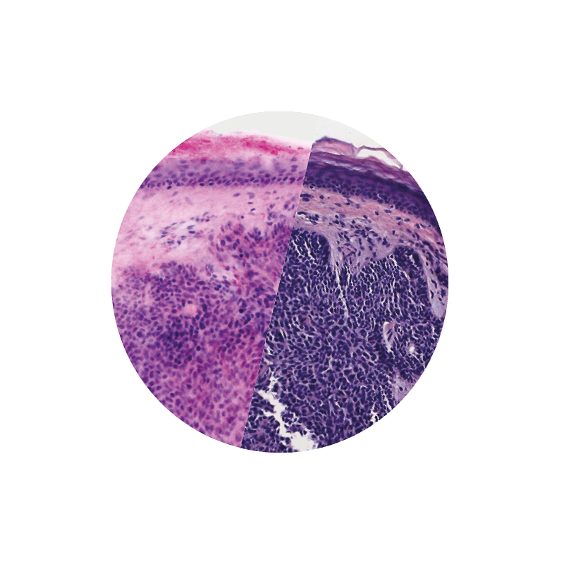
Images courtesy of Dr Javiera Pérez-Anker.
Basal cell carcinoma; imaged with the VivaScope 2500 (left) and after H&E staining (right)

Applications
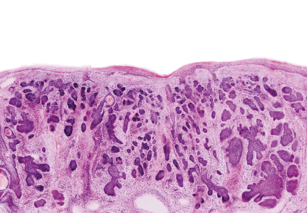
The VivaScope 2500 enables intraoperative assessment of tumor margins as well as immediate examination of biopsies. Surgical workflows and patient management can thus be significantly improved. The acquired images show subcellular details of the examined tissue.
100+ Publications
By using our technology of the VivaScope 2500, we are already part in more than 100+ publications. And there are more to come. The most application fields for instant digital pathology are so far Dermatology, Urology, EUS / EBUS – FNA / FNB, Organ Transplantation, Gastroenterology, Interventional Radiology and Senology / Gynaecology.


Find centers that already use the VivaScope 2500:
We are constantly expanding our clinic partners.
If you have any questions, contact us.

Seminars and trainings
We provide extensive and intensive on-site training to all of our customers. For a fast and precise application. You want a live demonstration or digital consulation? Than contact us any time.
Meet us - Congresses:
Take the opportunity to learn more about our products at the various trade shows and congresses around the world. Get in touch with our team on site.